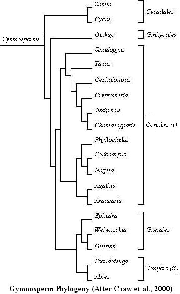
Gymnosperms are plants with seeds ( Spermaphytes) but with a naked ovule: the ovule is not included in a carpel that is an apomorphic feature of Angiosperms. A leaf-life structure (homologous to a leaf), a scale or megasporophyll bears the ovule. Gymnosperms are probably monophyletic and include: – Cycadales (Cycas, Zamia) that are the more primitive Gymnosperms (Basal Gymnosperms), – Ginkgoales, a sister-clade of the clade including Conifers and Gnetales, – Gnetales (Ephedra, Gnetum, Welwitschia) are monophyletic and nested within Conifers, – Gnetales are the sister-clade of Pinaceae. 
Fleshy fruit: berry or drupe
Fleshy fruits are characterised by a fleshy pericarp. It is usually the mesocarp that has high water content. A fleshy fruit is: – A berry when the endocarp is not lignified: grapes, tomato… Generally, berries have several seeds, rarely one: date, avocado… – A drupe or a stone fruit when the single seed, sometimes more, is surrounded by a stony layer (lignified endocarp): cherry, plum…
Dry fruit: follicle, capsule or achene
Dry and dehiscent fruits open at maturity permitting the release of enclosed seeds. A dry and dehiscent fruit is: – A follicle when it is many-seeded, formed from one carpel and dehiscing along one side only, – A pod or a legume when it is a follicle dehiscing along two sides, – A capsule when it is formed from more than one carpel. Dry and indehiscent fruits do not split at maturity to release seeds. It is the whole fruit that is dispersed by wind or insects. A dry and indehiscent fruit is: – An achene when it is indehiscent and 1-seeded with the seed coat distinct from the fruit wall (pericarp), – A samara when it is an achene with a flat wing formed from the pericarp, – A caryopsis when it is an achene with the seed coat (testa) united to the fruit wall. It is a feature of the grasses.
Angiosperm carpel
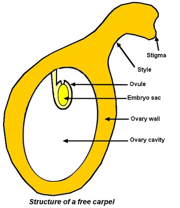
A good understanding of carpel organisation is imperative to analyse transformations that occur in an Angiosperm flower after fertilisation to obtain a fruit. A carpel is made of the 3 following parts: – a stigma, a wet surface with papillate cells, that is adapted for the reception and germination of pollen (see pages on pollen), – a style, more or less developed. The pollen tube grows between its cells during the pollen germination (see pages on pollen germination), – and, an ovary, the hollow and basal part of the carpel that contains inside its cavity an ovule (see pages on ovule). Inside ovule, there is the embryo sac (see pages on embryo sac).  Carpels can be free and gynaecium is then apocarpous. If ovaries are entirely fused together, gynaecium is syncarpous (see pages on syncarpous gynaecium). After fertilisation of the egg contained in the embryo sac (see pages on double fertilisation), many transformations occur: – the ovary develops into the fruit and the ovary wall gives the fruit wall (pericarp), – the ovules develop into the seeds and ovule integuments give seed coat, – the fertilised egg develops in the embryo that will be in the seed. Embryo will develop in a new plant during germination.
Double fertilisation in Angiosperms
When the pollen tube penetrates the embryo sac at the micropylar end, it goes through one synergid, burst and release the 2 sperm cells. One sperm cell and the egg fuse into a main zygote, diploid, that will give the seed embryo. The other sperm cell fuses with 2 well individualized or already fused polar nuclei. It gives a secondary zygote or endosperm that is triploid. It is called albumen in French to make the distinction with the Gymnosperms endosperm that is diploid tissue. Endosperm is usually triploid but its ploidy number can be different given that the number of polar nuclei can vary. Endosperm is a nutritive tissue which ends its development before that of the main zygote. When this last one will have ended its own development, endosperm has disappeared (seeds of Asteraceae or Fabaceae) or remains to go on its nutritive functions for the mature embryo during seed germination (seeds of Ricinus, seeds of Coco with a liquid endosperm).
